Collaborators
Edward A. Ross
(Division of Nephrology, Hypertension, and Transplantation)
Matthew J. Williams
(2Biomedical Engineering)
Takashi Hamazaki
(3Pathology, Immunology and Laboratory Medicine)
Marda Jorgensen
(Pathology, Immunology and Laboratory Medicine)
William L. Clapp
(Pathology, Immunology and Laboratory Medicine)
Gary W. Ellison
(4Small Animal Clinical Sciences)
Naohiro Terada
(Pathology, Immunology and Laboratory Medicine)
Introduction
The development of complex cellular scaffolds that facilitate the generation or repair of kidney-specific structures has challenged the tissue engineering field. Extracellular matrix (ECM) has organ-specific chemical composition and architectural arrangement, and has been shown to support cellular attachment, migration, and differentiation. In addition, ECM is biodegradable, and if properly extracted, has been shown in some models to have negligible immunogenicity.
Our team has previously produced intact cellular scaffolds through the decellularization of rat kidneys. The resultant organ-like structures retain the intricate vascular and tubular networks, allowing luminal delivery of cells through either arterial or ureteral pathways. Recently, the decellularization protocol has been further optimized to repeatedly produce scaffolds with only trace amounts of cellular debris. This significantly improved cellular response. Ultimately, it is thought that such an elaborate structure could be repopulated ex vivo with appropriate cells and implanted into those in need of kidney transplantation.
Embryonic stem (ES) cells have been shown to differentiate into cells specific to the adult kidney. Also, organ-specific ECM substrates have been shown to direct ES cell differentiation. Therefore, as a first pass, we have seeded the scaffold with murine (B5/GFP+) ES cells. These cells allow fluorescent tracking and will show if the produced scaffolds effect cell differentiation, without the addition of lineage-inducing culture additives. These experiments will show the quality of cell dispersion through arterial injection, succeeding proliferation and migration patterns, and resultant cellular morphologies and differentiation tendencies.
MATERIALS AND METHODS
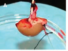
- Rat kidneys were harvested using strict IACUC care guidelines. The renal arteries were cannulated to allow attachment to the perfusion-based decellularization system. The kidneys were decellularized over 5 days using successive arterial perfusions of heparinized saline, vasodilators, Triton X100, salts, DNAse, sodium dodecyl sulfate (SDS), and distilled water (modified from K. Brendel, 1978). The resultant scaffold structure was imaged by light microscopy, and conservation of key basement proteins was validated with collagen IV and laminin immunohistochemistry (ABCAM). All immunohistochemical stains were developed with DAB and counterstained with hematoxylin.
- Suspensions of 2 million murine pluripotential embryonic stem (ES) cells (B5/GFP+) were injected into the whole organ via arterial cannulation. The ES cells were prepared through trypsinization and suspension in IMDM, supplemented with 20% fetal bovine serum (Atlanta Biologicals), 2 mM L-glutamine, 100 units/ml penicillin, 100 μg/ml streptomycin (GIBCO BRL), 0.25 μg/ml Amphotericin B (Cellgro) and 300 μM monothioglycerol (Sigma).
- After a day to allow for cell adherence, the whole organs were cut into thick transverse sections and incubated up to 10 days in a multi-well dish. Cell distribution and migration and expansion patterns were tracked through GFP fluorescence imaging.
- Evaluation of scaffold cultures for resultant cell morphologies and differentiation patterns included H&E histochemistry and various immunohistochemical stains including Pax-2 (Zymed), Pancytokeratin (Ventana), and KSP-cadherin (Zymed).
ORGAN DECELLULARIZATION
Our decellularization process, which uses an arterial perfusion apparatus, produced whole-organ kidney cellular scaffolds. These ECM scaffolds were blanched, translucent, and spongy. In addition, these scaffolds showed preservation of vascular and collecting system networks, facilitating cell delivery through either anterograde or retrograde pathways.
The basement membranes of the tubules and glomeruli appeared undamaged with only trace evidence of cellular debris. An older decellularization process, utilizing the ionic detergent sodium deoxycholate as opposed to sodium dodecyl sulfate (SDS), shows a stark contrast with the new protocol. H&E histochemistry, collagen IV and laminin immunohistochemistry confirmed removal of rat cells and continuous, intact basement membrane structures.
CELL SEEDING
Implanted murine GFP+ ES cells, delivered through the renal artery, showed extensive glomerular localization. These cells, over time, expanded into the adjacent vessels, including the tubular interstitium, and descended into the medulla. It is likely that most of the cells remained confined to the vascular networks, however, histochemistry suggests some cells have escaped into the tubular basement membranes.
Cultured cells showed marked patterns of proliferation and exhibited distinct location-dependent morphologies. A survey of noteworthy areas shows cells appear to be responding to the various basement membranes. Cells adjacent to basement membranes, notably vascular walls, are less apoptotic then those in the luminal spaces.
DIFFERENTIATION ANALYSIS
While more extensive analyses are underway, results indicate potential in the produced scaffold to effect stem differentiation of injected ES cells.
Pax-2, a key protein for kidney development, appears in cells seeded in the scaffolds as early as 3 days post injection. Throughout the culture durations, pax-2 positive cells are present in abundance. KSP-cadherin, which also plays a role in development, is mostly absent from cultured cells, with the exception shown. Preliminary assessments of WT-1, another protein in kidney development; Oct-4, an ES cell marker; and cytokeratins, a general marker of epithelial differentiation, has been performed. As of yet there is no WT-1 expression, extensive clusters of cytokeratin expression, and conflicting results (yet to be resolved) on OCT-4 patterns. All proteins are absent in the unseeded scaffold, and all but OCT-4 are absent in the ES cells before injection.
CONCLUSIONS
Whole kidneys can be decellularized using an arterial perfusion protocol. The optimized protocol we have developed repeatedly produces complex ECM scaffolds with negligible cellular debris, retention of laminin and collagen IV, and preservation of the intricate vascular, glomerular, and tubular architectures.
Murine embryonic stem cells, seeded through the renal artery, showed extensive glomerular distribution in the rat-derived scaffolds. The ES cells, cultured up to 10 days, revealed distinct, scaffold-dependent patterns of proliferation, migration, changed cell morphology, and differentiation.
Ultimately, to further evaluate kidney ECM as a scaffold, and isolate the various factors influencing cellular response, more studies need to be conducted. Analysis of proliferation and apoptosis patterns through KI-67 and CC3 immunohistochemistry is currently ongoing. In addition, studies are being pursued to compare the cellular morphologies, expression patterns, and expression levels of implanted cells as compared to embryoid bodies (EBs). Embryoid bodies are ES cells that are allowed to aggregate and differentiate outside the influence of a scaffold. Some proteins, such as pax-2 and cytokeratins, are also expressed in EB culture, and therefore it remains to be seen if the levels and patterns of expression are changed when cultured in the scaffold. Finally, ureteral seeding is being successfully pursued. This alternate seeding technique will expose the implanted cells to the tubular and collecting ductular basement membrane of the kidneys.